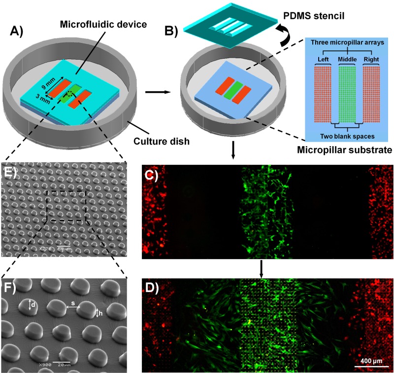
FIG. 1.
Schematic illustration of the experiment procedures and characterization of the microfluidic device. (a) The microfluidic device used in the current study, which was composed of four layers, a PDMS stencil, a PDMS micropillar substrate, a thin PDMS membrane (not shown), and a polystyrene culture dish. The PDMS stencil was sealed to the micropillar substrate, and each type of cells was seeded into the appropriate region (from left to right, HaCaT, ESF-1, and HUVEC cells, respectively). (b) The PDMS stencil was peeled off after the cells were attached and cultured on the micropillar substrate for 24 h. (c) A typical fluorescence image of well-distributed cells after the stencil was just peeled off. To visualize clearly the coexistence of cells, HaCaT (left), ESF-1 (middle), and HUVEC (right) cells were specifically stained using CellTracker Orange, CellTracker Green, and CellTracker Orange, respectively. (d) A typical fluorescence image of cells after wounding, corresponding to 36 h. (e) SEM image of the micropillar substrate. (f) Enlarged image of the square in the dotted lines in (e).

