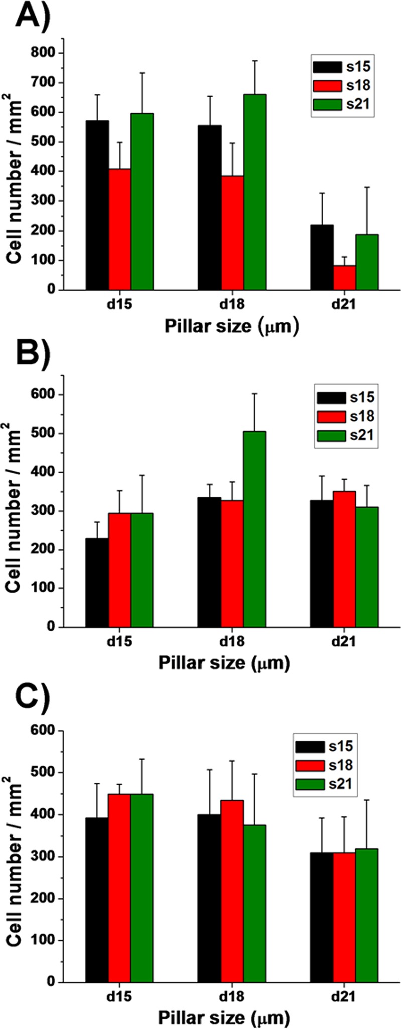
FIG. 2.
Quantitative assessment of the cell attachment on the different micropillar substrates. Histograms present cell numbers of (a) HaCaT, (b) ESF-1, and (c) HUVEC cells. Cell numbers were obtained by manual counting of AO stained cells. For clarity, the different diameters (15, 18, and 21 μm) and spacing (15, 18, and 21 μm) of the micropillars were denoted as d15, d18, and 21 μm and s5, s18 and s21 μm, respectively.

