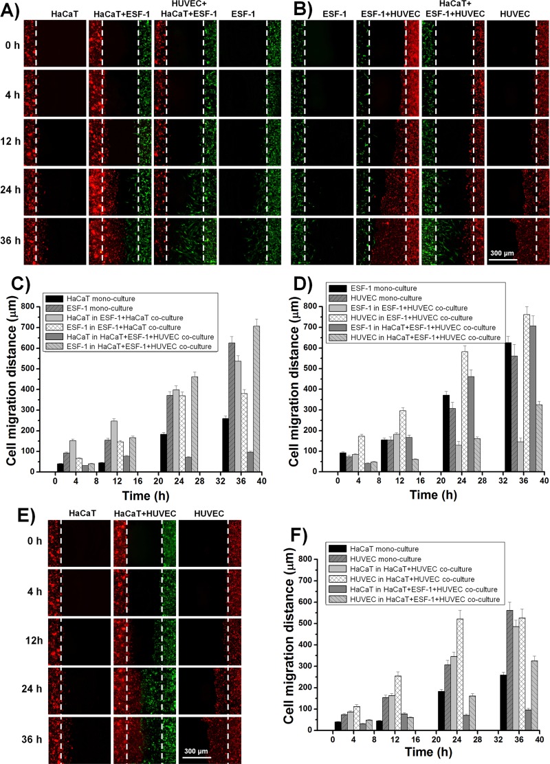
FIG. 6.
Dynamic cell migration under different conditions after the wound. (a) Dynamic migration of HaCaT and ESF-1 cells in their mono-culture and co-culture. (b) Real-time migration of ESF-1 and HUVEC cells in their mono-culture and co-culture. (e) Dynamic migration of HaCaT and HUVEC cells in their mono-culture and co-culture. (c), (d), and (f) Quantitative analysis of cell migration distances during (a), (b), and (e). The initial leading edges (shown as white dashed lines) represent baselines for the migration assay.

