Fig. 1.
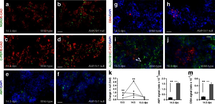
ALDH1B1 controls timing of commitment and proliferation of embryonic pancreas progenitors and is expressed in nascent beta cells. (a–h) The number of NGN3+ (a, b), C-PEP+ (c, d), AMY+ (e, f) and DBA+ (g, h) cells was increased in pancreases of Aldh1b1 tm1lacZ null mice compared with wild-type mice. (i, j) Co-expression of ALDH1B1 with newly formed beta cells remains until 15.5 dpc (arrows) but is lost at 16.5 dpc. (k) Quantification of C-PEP+ (squares), NGN3+ (triangles) signal (DAPI normalised) and number of PH3+ epithelial (diamonds) cells per islet at 13.5, 14.5 and 15.5. dpc showed a transient increase (expressed as fold increase compared with wild-type) in Aldh1b1 tm1lacZ null pancreases. Comparisons were done for the same day. (l, m) Quantification of AMY+ (l) and DBA+ (m) signal (DAPI normalised) at 14.5 dpc showed a strong increase (expressed as ratio over wild-type) in the Aldh1b1 tm1lacZ null pancreases (black bars, wild-type; grey bars, null). Results are from four to six embryos per genotype and per embryonic stage, and values are representative of eight sections per embryonic pancreas spanning the entire organ. *p < 0.05 and **p < 0.01. Scale bars, 50 μm (a, b), 100 μm (c, d), 20 μm (e–h) and 15 μm (i, j). E-CAD, E-cadherin

