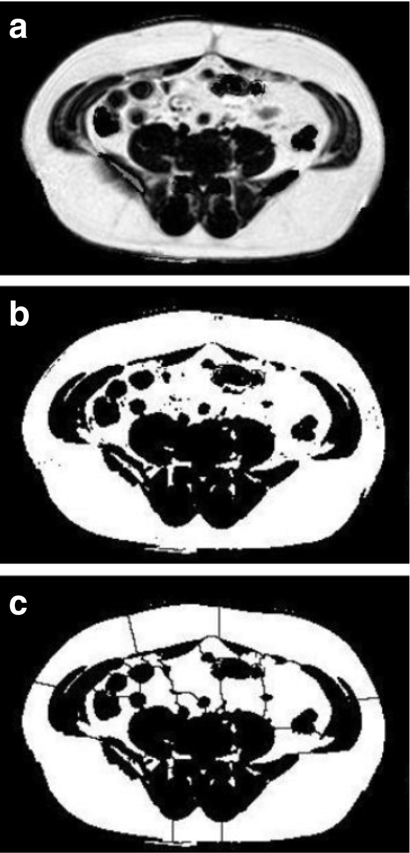
Fig. 3.
Quantifying visceral fat. (a) The three-point Dixon fat fraction map acquired at the L4–L5 junction. (b) Binary thresholding of structures containing more than 50% fat (visceral and subcutaneous fat) from those with less. Total area is evaluated. (c) Application of thresholding algorithm divides segmented image into chunks and separates visceral and subcutaneous fat around the boundary of the chest wall. Selection of subcutaneous fat and any external signals allows measurement of this area and subtraction from the total to yield visceral fat area

