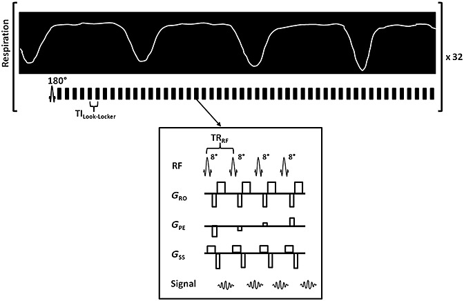
Figure 1.

Schematic diagram of the respiratory-triggered, Look–Locker T1 mapping sequence with a spoiled, single-slice, gradient-echo readout. The inversion pulse is end-expiration triggered, followed by a free-breathing, segmented, Look–Locker sampling train. Each segmented block is separated by TILook–Locker; each block contains four sampling pulses with TRRF. The sequence is performed twice with differing inversion slice thicknesses as part of the flow-sensitive alternating inversion recovery-arterial spin labelling (FAIR-ASL) design; a hepatic ASL (HASL) dataset is completed in 15 min.
