
Figure 2.
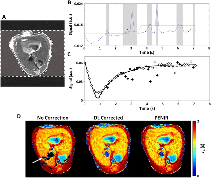
The phase-encoded noise-based image rejection (PENIR) scheme for retrospective removal of motion-corrupted data. (A) Axial image of a mouse liver with an example user-determined region of interest (ROI) (broken line) of the extracorporeal area in the phase-encoding direction. (B) Significant increases in the mean noise signal (blue line) above a signal-generated threshold (red line) will result in image omission (shaded areas). (C) Inversion recovery fitting in a liver region will typically overestimate T1 without correction (broken line) compared with rejection (full line) because of reduced tissue signal from corrupted images (filled diamonds). (D) A comparison of T1 maps shows that, with no correction, the respiration artefacts may lead to unsuccessful fitting and generate areas of signal dropout (arrow). With both data-logger (DL) and PENIR retrospective gating, these areas of dropout are recovered – no significant differences in mean T1 were measured between these two retrospective conditioning modes (t-test, p > 0.05).