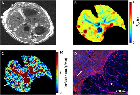
Figure 5.

Application of hepatic arterial spin labelling (HASL) to a model of liver metastasis. (A) High-resolution T2-weighted fast spin echo images show the tumours (arrow) as hyperintense relative to the liver tissue (outlined). (B) In the corresponding slice-selective T1 map, the metastases can be delineated by a raised T1 (2.24 ± 0.54 s) relative to liver tissue. (C) Across n = 6 mice, a significant reduction in perfusion (outlined) was measured within the metastases (1.1 ± 0.5 mL/g/min, mean ± standard deviation) relative to the liver tissue. This difference in perfusion measured by HASL was confirmed by histology. (D) The fluorescence image shows a section of normal liver containing sinusoid vessels (arrow), demarcated from an adjacent SW1222 colorectal liver metastasis (star) by a large reduction in the presence of blood vessels. Vascular structures are shown in red (anti-CD31 antibody) and perfusion in blue (by the injected marker Hoechst 33342).
