Fig. 4.

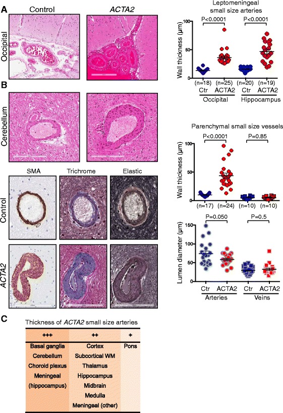
Small-size artery disease in ARCD. a. H&E of leptomeningeal vessels overlaying the calcarine cortex. The graph shows the distribution and mean ± SEM of the transmural thickness for leptomeningeal small arteries from two brain regions. b. Analysis of parenchymal small vessels from the cerebellum shows thickened walls with characteristic SMC proliferation and fibrosis. The quntifications of transmural wall thickness and lumen diameter are shown for both small arteries and veins. The scoring of lumen diameters was performed on 20 α-SMA labeled vessels in each category. Control and mutant groups for each category were compared by t-test. c. Distribution of the ACTA2 mutant small size arteries with variable wall thickness across brain regions. +++, ++ and + represent 2.5–4, 1.5–2.5 and <1.5 fold increase vs. control
