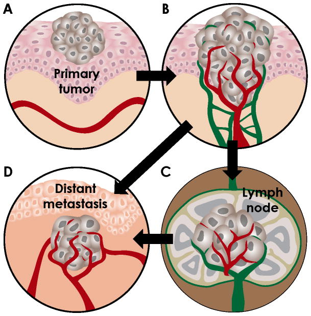
Figure 1. Illustration of steps in the metastasis process.
A. Early carcinomas are confined to the epithelial compartment and receive their oxygen and nutrients by diffusion. B. To grow beyond 1mm3, tumors acquire neovascularization. Increased tumor-associated vascular and lymphatic density increases the propensity for tumor dissemination. Blood vessel, red; lymphatic vessel, green. C. Tumor cells can escape via lymphatic vessels and arrest in sentinel lymph nodes. Tumor cells in the lymph node may invade local blood vessels or remain in the lymphatic system to be recycled to the vascular system. D. Tumor cells may also invade blood vessels in the tumor (intravasation), travel in the circulation and exit in the new organ environment (extravasation). Tumor expansion again requires angiogenesis in the secondary site. Tumor cells can metastasize via the vascular system (B→D) or the lymphatic system (B→C→D).

