
Figure 1.
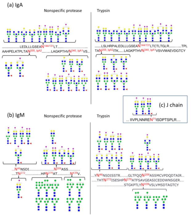
Site-specific glycan mapping for IgA an IgM using specfic and non-specfic proteases. (a) Glycan map for IgA. IgA1 glycosite N144 and IgA2 glycosite N131 share the same tryptic peptide, thus cannot be distinguished from each other. While no fucosylation were observed for glycosite N144/131, glycans on glycosite N205 (IgA2) and N340 (IgA2) are all fucosylated. (b) Glycan map for IgM. Glycans on site N46, N209 and N272 are all highly sialylated, while mostly high mannose type glycans were observed on site N279 (one hybrid-type glycan identified) and N439. (c) Three glycans were observed for J chain. Symbol key: yellow circles = galactose (Gal); green circles = mannose (Man); blank circles = hexose (Hex); blue squares = N-acetylglucosamine (GlcNAc); yellow squares = Nacetylgalactosamine (GalNAc); red triangles = fucose (Fuc); purple diamonds = Nacetylneuraminic acid (Neu5Ac).