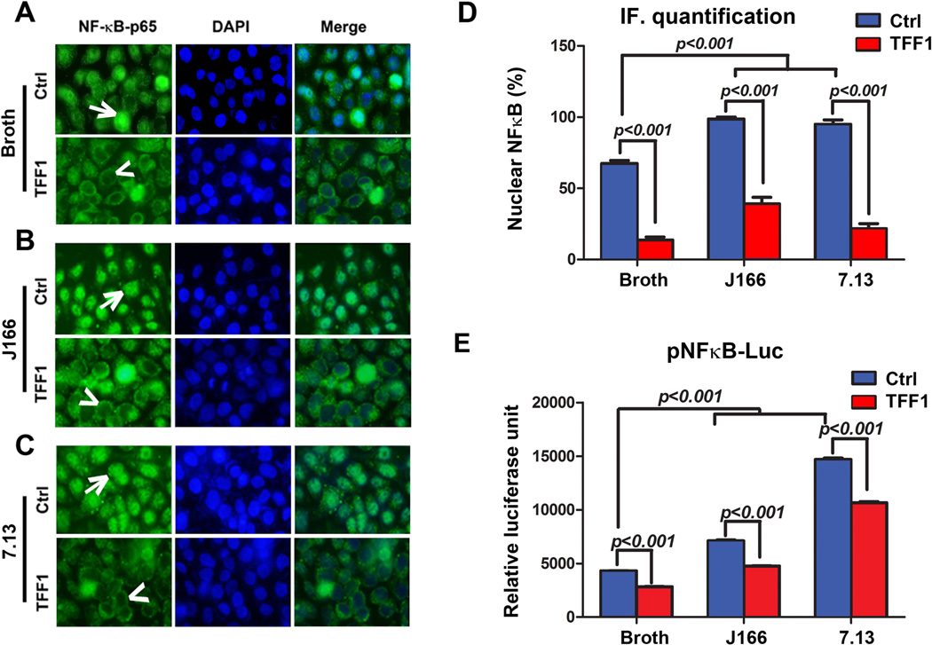
Figure 1. TFF1 reconstitution alters H. pylori-mediated nuclear translocation and transcriptional activation of NFκB.
A–C) In vitro immunofluorescence assay of NFκB-p65 in uninfected and H. pylori-infected AGS cells stably expressing pcDNA or TFF1 indicating nuclear localization of NFκB-p65 (green fluorescence, arrows) in AGS-pcDNA cells and absence of nuclear NFκB-p65 staining (arrowheads) in AGS-TFF1 cells. (A) Uninfected cells (Broth). (B) Cells infected with H. pylori J166 strain. (C) Cells infected with 7.13 strain. DAPI (blue) was used as a nuclear counterstain. Original magnification, ×40. D) Graph shows the quantification of nuclear NFκB-p65–positive staining in at least 200 counted cells, indicating an increase of NFκB-p65 nuclear staining in AGS-pcDNA cells, which decreases in AGS-TFF1 cells after infection. Results presented as percentage ± SEM. E) The luciferase reporter assay using a pNFκB-Luc reporter plasmid. H. pylori infection of AGS-pcDNA cells significantly increased the luciferase activity, which was reduced after reconstitution of TFF1. The bar graphs represent the mean ± SEM of 3 independent experiments.

