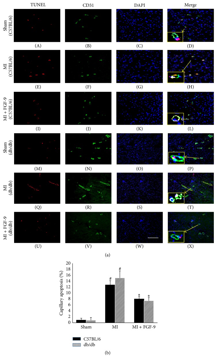
Figure 2.
Effects of FGF-9 on capillary apoptosis following MI in C57BL/6 and db/db mice. Representative images demonstrating capillary apoptosis are illustrated in (a) for C57BL/6 (A–L) and db/db (M–X) mice with TUNEL positive nuclei in red (A, E, I, M, Q, and U), CD31+ve cells in green (B, F, J, N, R, and V), total nuclei stained blue with DAPI (C, G, K, O, S, and W), and merged images (D, H, L, P, T, and X). The smaller boxes (D, H, L, P, T, and X) are enhanced images shown to illustrate colocalization of TUNEL, CD31, and DAPI within a single vessel. Scale bar = 100 μm. (b) Quantitative data suggest FGF-9 inhibits capillary apoptosis following MI in C57BL/6 and db/db mice. # p < 0.05 versus sham and ∗ p < 0.05 versus MI.

