Figure 3.

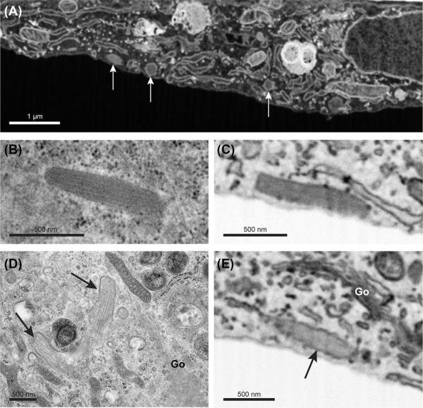
Weibel–Palade bodies imaged by SEM and TEM. Weibel–Palade bodies (WPBs) imaged by SEM and TEM in endothelial cells. (A) SBF-SEM slice showing cross-sectioned WPBs (arrows). (B) Longitudinal-sectioned WPB imaged by TEM displaying internal striations of Von Willebrand factor tubules. (C) Inverted SEM image of a longitudinal-sectioned WPB that displays a uniform stained interior. (D) Immature WPBs (arrows) near the Golgi apparatus (Go) imaged by TEM. Separate VWF tubules are clearly visible. (E) Inverted SEM image of an immature WPB near the Golgi apparatus (Go). Interior is less electron dense than the WPB in panel C but the VWF tubules are not resolved. Scale bar panel (A) is 1 μm. Scale bar panels (B)–(E) is 500 nm.
