Figure 4.

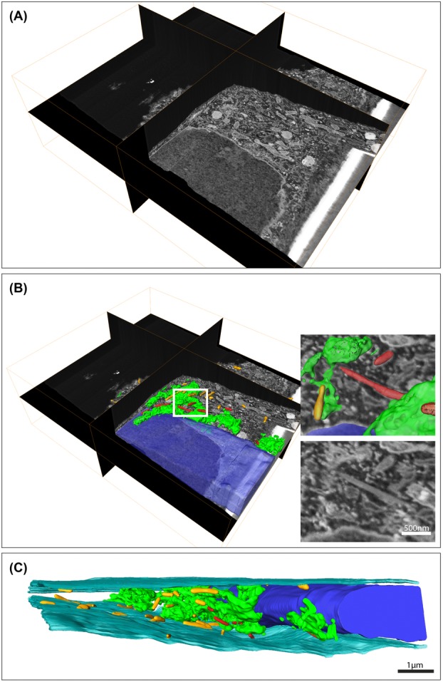
Serial block face (SBF)-scanning electron microscopy (SEM) reveals Weibel–Palade bodies in close association with the Golgi. Analysis and modelling of a large SBF-SEM stack to study WPBs in relation to the Golgi apparatus in endothelial cells. (A) Three-dimensional overview of the data set. The data set consist of almost 1600 images and was acquired at 25 000× magnification using a 10 nm slice thickness. For processing, the data were binned two times in x and y which resulted in a voxel resolution of 7.4 × 7.4 × 10 nm. (B) Segmentation of the volume reveals the large size of the Golgi apparatus (green) and the WPBs (red) that are in close association with the Golgi. In addition, we segmented the peripheral WPBs (yellow) and the nucleus (blue). The inserts show one of the WPBs that was found in close relationship with the Golgi. Scale bar is 500 nm. To have a better view on the ultrastructure, the data were rotated with respect to the orientation in which the data were acquired for panels (A) and (B). (C) Segmentation of the cell membrane additionally reveals the confined and flat morphology of the endothelial cell. Scale bar is 1 μm.
