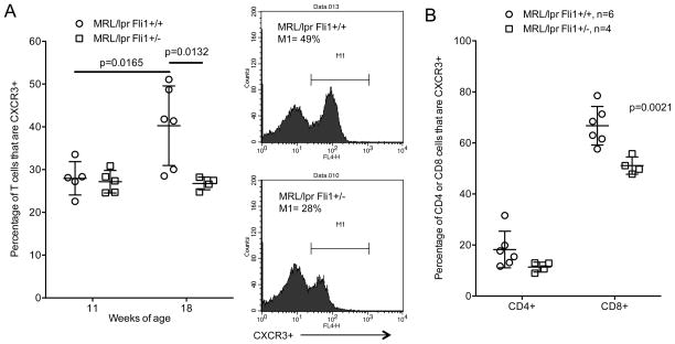
Fig. 4.
The percentages of CXCR3+ T cells are significantly decreased in 18 week-old MRL/lpr Fli1+/− mice. A) Percentages of CXCR3+ cells were quantified by flow cytometry in T cells negatively isolated from spleens of 11 and 18 week-old MRL/lpr Fli1+/+ and Fli1+/− mice (n=4–6 for each group. Example of flow data is shown to the right of the graph. B) Percentages of CD4+ and CD8+ T cells that are CXCR3+ in the 18 week-old samples analyzed in (A).

