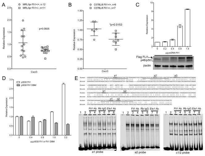
Fig. 6.
FLI1 regulates Cxcr3 expression in T cells. Cxcr3 message levels were measured by real-time PCR in reverse-transcribed RNA from negatively isolated splenic T cells of 16–18 week-old MRL/lpr (A) and C57BL/6 (B) Fli1+/+ and Fli1+/− mice. C) The Jurkat T cell line was transfected with a CXCR3 promoter/reporter vector containing the human CXCR3 (hCXCR3) promoter driving luciferase expression and increasing amounts of the FLI1 expression vector pcDNA Flag FLI1 (Flag FLI1). Cells were stimulated for 24 hours and whole cell extracts were analyzed for luciferase expression and subjected to western immunoblotting with antibodies to FLI1 and βactin (below graph). D) The Jurkat T cell line was transfected as indicated in (C) with the hCXCR3 promoter/reporter vector, but with increasing amounts of the FLI1 expression vector pSG5 FLI1 or the FLI1 DNA binding mutant (DBM) expression vector pSG5 FLI1 DBM. Transfections were performed at least three times with similar results. Representative experiments are presented. E) In vitro DNA binding assays were performed with the indicated hCXCR3 probes containing putative conserved ETS binding sites and nuclear extract from stimulated Jurkat T cells without or with anti-FLI1 or anti-ETS1 antibodies or normal IgG. Aligned sequences of the human and mouse CXCR3 promoters with locations of probes and putative ETS sites are shown above the EMSA blots.

