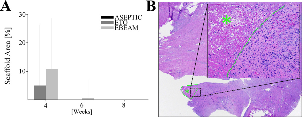
Figure 3. Scaffold resorption.
A - Percentage of ACL sections covered by scaffold material at 4, 6, and 8 weeks. No statistically significant difference between EBEAM and ETO after 4/6 weeks. At 8 weeks there were no scaffold residuals left in any group. Aseptic group was only tested at 8 weeks and did not show any scaffold residuals. B - H&E stained ACL section from EBEAM group after 6 weeks (20× magnification) with scaffold residual (green outline/star). Inset showing scaffold area (200× magnification), border between scaffold area (green star) and adjacent connective tissue green. Scaffold material characterized as acellular eosinophilic material. The scaffold material was not associated with significant inflammation or hemorrhage but did have some fibrous ingrowth.

