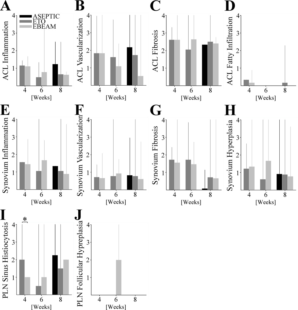
Figure 5. Histological Scoring of Anterior Cruciate Ligament (ACL), Synovium, and Popliteal Lymph Node (PLN).
Barplots indicating mean and spikes 95% CI of A - ACL inflammation, B – ACL neovascularization, C - ACL fibrosis, and D - ACL fatty infiltration, as well as E - synovial inflammation, F - synovial neovascularization, G - synovial fibrosis, and H - synovial hypertrophy, and also I - popliteal lymph node sinus histiocytosis (* indicating statistical significant difference with P< .001 and large effect size of d = 1.41) and J - popliteal lymph node follicular hyperplasia at 4, 6, and 8 weeks for ETO and EBEAM and at 8 weeks for ASEPTIC group.

