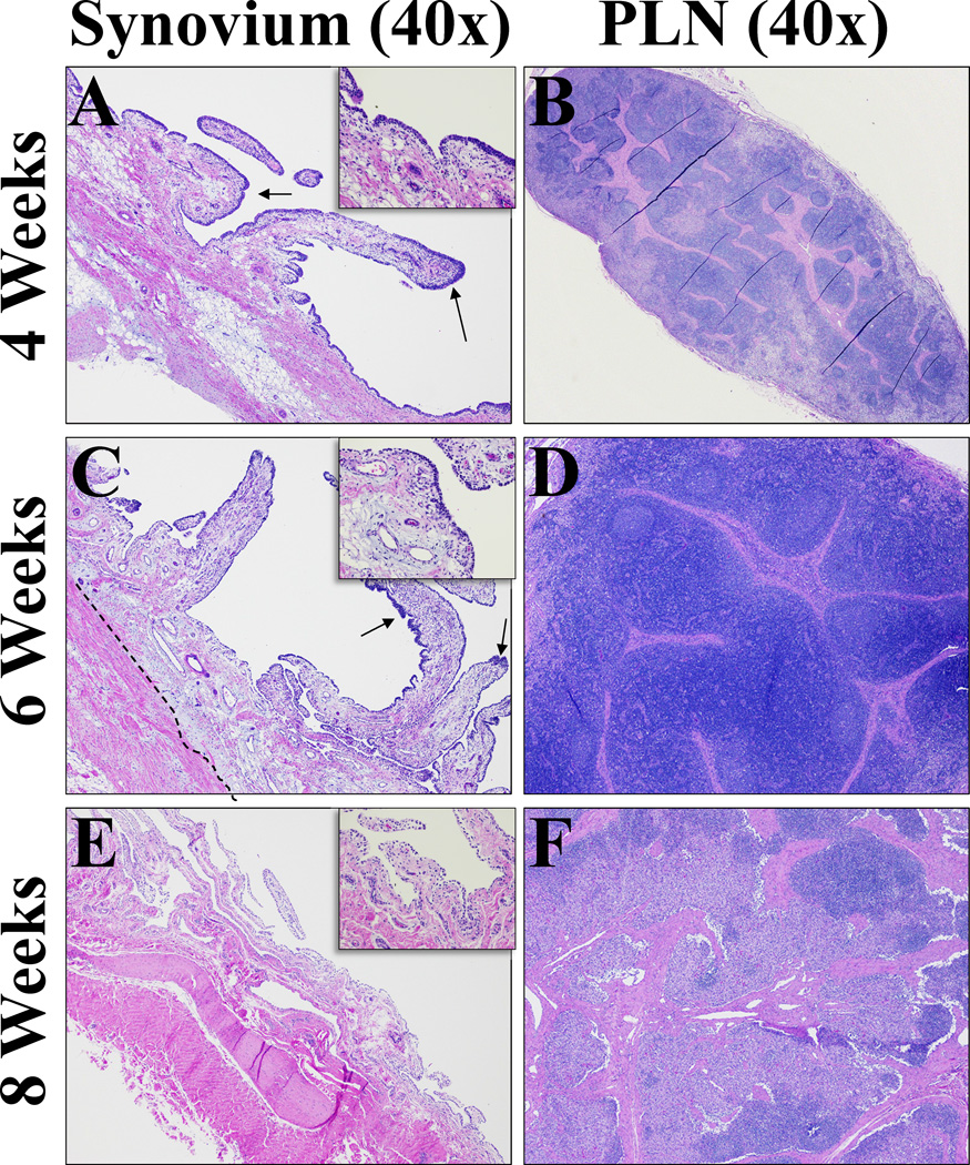
Figure 6. Histological sections from synovium and popliteal lymph node (PLN) representative of all treatment groups.
A - H&E stained synovium section after 4 weeks (40× magnification). Diffusely thickened intima, multifocal areas of prominent hyperplasia (arrows). Subintimal inflammation present consistent with ongoing healing. Fibrosis slightly increased. B - H&E stained popliteal lymph node section after 4 weeks (40× magnification). Prominent sinus histiocytosis consistent with mild lymphadenopathy as a result of draining inflammation from implant site, not considered an adverse reaction. C - H&E stained synovium section after 6 weeks (40× magnification).Still multifocal areas of hyperplasia (arrows), but overall intima thin and lined by 1–2 cells as seen in control tissues. Subintimal fibrosis remained increased (dashed line). D - H&E stained popliteal lymph node after 6 weeks (40× magnification). Mild follicular and paracortical and sinus histiocytosis. E - H&E stained synovium section after 8 weeks (40× magnification). Resolution of intimal hyperplasia, subintimal fibrosis and inflammation. Collagen fibers approach thin intimal layer.F - H&E stained popliteal lymph node section at 8 weeks after bio-enhanced ACL repair 40× magnification for a general view. Follicular and paracortical and sinus histiocytosis continues to resolve.

