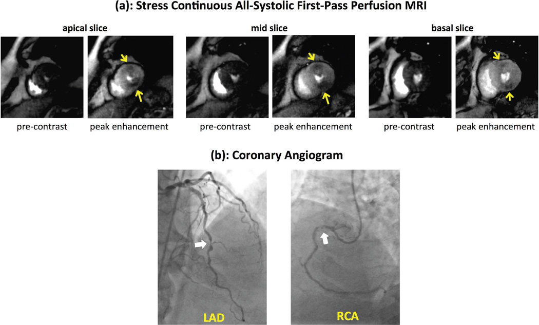
Figure 9.
(a): Adenosine stress myocardial perfusion MR images at the apical, mid, and basal ventricular levels, and (b) coronary angiogram performed 3 weeks after the MRI study in a second CAD patient. MRI was performed using the proposed all-systolic non-ECG-gated approach. For each slice in (a), the left panel shows pre-contrast phase for the stress scan and the right panel shows the peak myocardial enhancement phase (no rest study was performed). The observed stress perfusion defects, highlighted by yellow arrows, are consistent with the angiographic images in (b), which show significant stenoses (≈70%) in mid LAD and a high-grade stenosis (≈90%) in proximal RCA (dominant vessel). All images are of high quality, specifically in terms of the myocardial contrast between hypoperfused vs. normal territories, and no subendocardial dark-rim artifact is seen.

