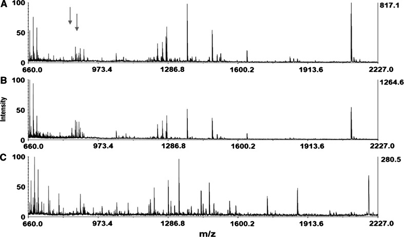
Fig. 3.
MALDI-TOF-MS analysis of three single retrograde-stained neurons containing no heptapeptides and located in different regions of the same right parietal ganglion (nonHP neurons). Note the total overlap in peptide content of the two spectra shown in A and B and the mostly differential expression of neuropeptides in spectrum C. The arrows indicate the predicted place for the two dominant heptapeptides (S/GDPFLRFamide) absent in these neurons (color figure online)

