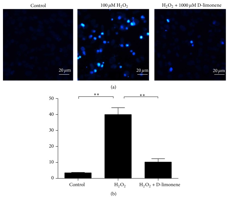
Figure 3.
Protective effect of D-limonene against H2O2–induced apoptosis. HLECs were pretreated with 1000 μM D-limonene for 12 h and then treated with 100 μM H2O2 for 24 h. The cells were then stained with Hoechst 33342 and observed using a fluorescence microscope. (a) Morphological apoptosis was observed using an inverted microscope. Cells that were exposed to H2O2 exhibited morphological changes typical of apoptosis, such as chromatin condensation and nuclear shrinkage. (b) The statistical analysis of the nuclear morphological change is shown. At least 70 cells in three different fields were counted per well in three separate experiments. ∗∗ P < 0.01.

