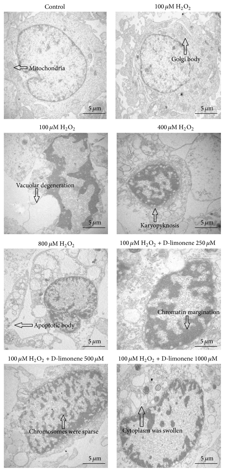
Figure 5.

HLECs were photographed by an electronic microscope. Cells that were exposed to H2O2 (100, 400, and 800 μM) for 24 h exhibited morphologic changes typical of apoptosis, such as cell shrinkage, irregular nuclear outline, chromatin condensation, apoptotic body, and cytoplasm vacuolization. In the D-limonene-pretreated HLECs, the cellular ultrastructure appeared to be more improved than that of cells treated with 100 μM H2O2 alone.
