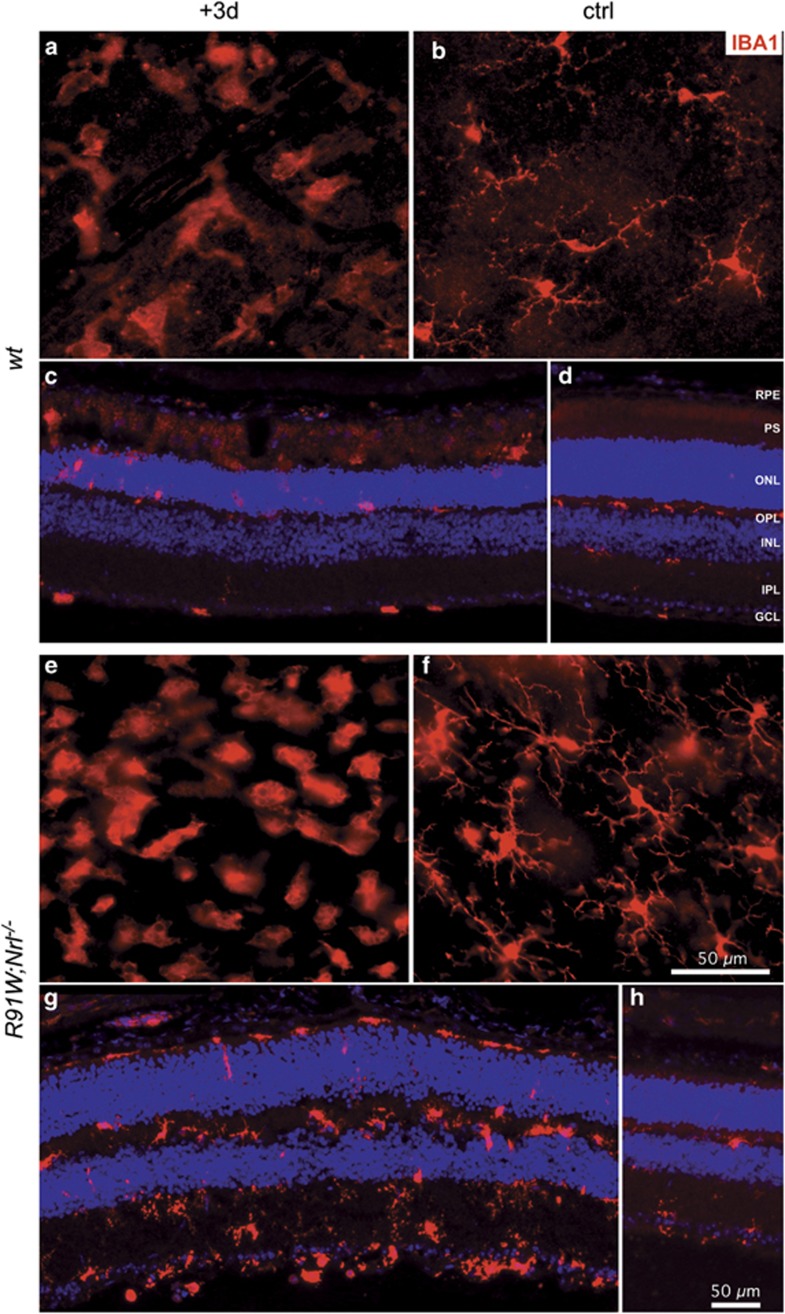
Figure 3.
Microglia/macrophage accumulation within the hotspot area at 3 days (+3d; a, c, e, g) following BLD in wt and R91W;Nrl−/− retinas. Undamaged regions outside the hotspot served as internal controls (ctrl; b, d, f, h). Shown are retinal flat mounts (a, b, e, f; the focal plane in the outer plexiform layer) or retinal cryosections (c, d, g, h) immunostained for IBA1. RPE: retinal pigment epithelium; OPL: outer plexiform layer; IPL: inner plexiform layer; other abbreviations as in Figure 1. N=3. Scale bars as indicated

