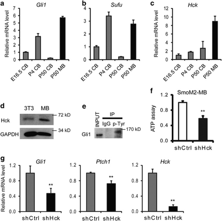
Figure 6.
Hck is highly expressed in SmoM2-induced medulloblastoma cells and required for its growth. (a–c) Expression levels of (a) Gli1, (b) Sufu and (c) Hck in cerebellum (CB) during different developmental stages and in SmoM2-induced medulloblastoma (MB) are shown. (d) The levels of Hck protein in NIH3T3 cells and in SmoM2 medulloblastoma were determined by western blot. (e) Endogenous Gli1 protein in medulloblastoma is Tyr phosphorylated. SmoM2 medulloblastoma lysates were precipitated with 4G10 antibody against phospho-Tyr and probed by western blot using antibodies against Gli1. (f) Cultured SmoM2 medulloblastoma cells were infected with lentiviruses expressing control (scrambled shRNA) or shHck. The survival rates of Hck-deficient tumor cells relative to the control cultures were measured using an ATP cell viability assay. (g) RT–PCR analyses of the expression levels of Gli1, Ptch1 and Hck in cultured SmoM2 medulloblastoma cells with Hck RNAi knockdown. Presented are means plus s.d.; n=3. Significance was determined by Student's t-test; **P<0.01.

