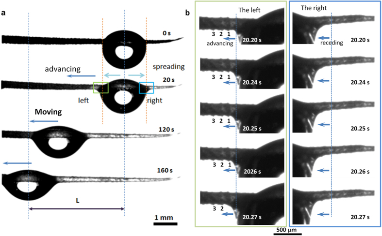
Figure 3.
(a) Drop transport on the scale-covered spine. A drop (2 μl) was deposited on horizontal spine. After ~20 s, the drop spread asymmetrically along the spine. Subsequently, the drop moved directionally at ~120 s. After ~160 s, drop almost moved along the spine. (b) In-situ observation of drop moved from one scale to another (from ~20.20 s to ~20.27 s). The left-side images show the liquid of the drop moved along the scales and advancing to cover over scale 1 at ~20.27 s. The right-side images show the right-hand of the drop moved along the scales, receding to show the scale at ~20.27 s.

