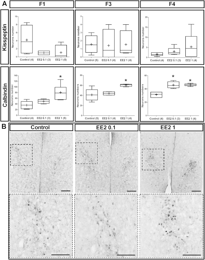
Figure 4. Unlike kisspeptin neurons, developmental EE2 exposure disturbed sexually dimorphic calbindin immunoreactive neurons in the medial PreOptic Area (mPOA).
(A) Tukey’s boxplots of the number of kisspeptin and calbindin-immunoreactive neurons per brain section in mPOA of F1, F3 and F4 males. The line in the middle of the box is plotted at the median and (+) is the mean. Numbers in brackets represent litter numbers. Kruskal-Wallis test with Dunn’s post-test with *p-value < 0.05. (B) Representative anti-calbindin immunostaining in the mPOA showing the location of the sexually dimorphic calbindin population (NDS-POA). Images of the first panel present a low magnification giving an overview of the location of NDS-POA broadly delimited by the dotted squares magnified in the panel below. Scale bars = 100 μm.

