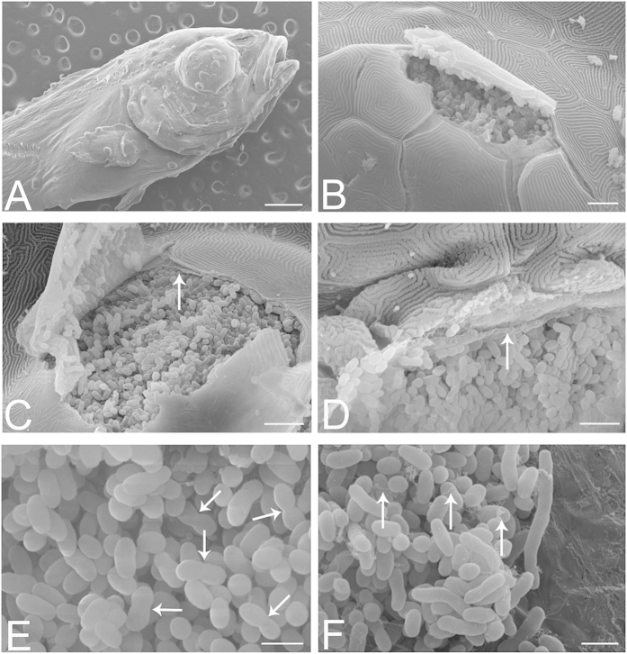
Figure 3. SEM of infected larvae at 21 dph.
(A) Surface view of larva, showing cysts on skin and fins. (B) A ruptured cyst beneath the skin appears to be densely packed with bacteria. (C) Ruptured cyst revealing the bacteria to be separated from the epithelial cells by a membrane (arrow) with a fibrous-like appearance, and with multiple fenestrations or perforations. Should the cyst microenvironment be tightly regulated by the enveloping cells, then presumably these would need to form tight junctional contacts with each other and with the cyst itself. (D) Bacteria appear to be in intimate contact with the surrounding membrane (arrow), which appears to partially fold around the bacteria, creating shallow bays for them. (E) Within a cyst, bacteria appear to be dividing (arrows). (F) Another view shows elongated bacterial forms and filaments (arrows). Scale bars represent: (A) 500 μm, (B) 10 μm, (C,D) 5 μm, (E,F) 2 μm.

