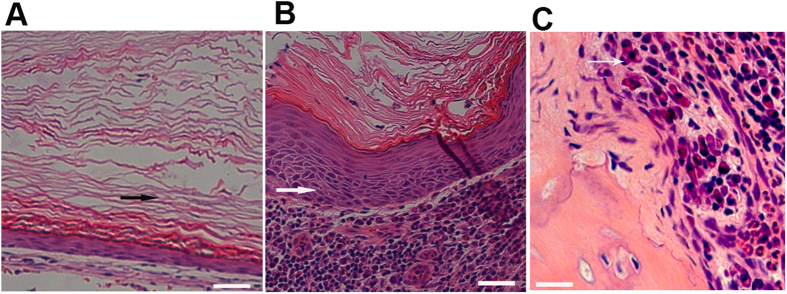
Figure 1. Pathological profiles of congenital versus acquired cholesteatoma and eroded bone.
Representative serial sections of cholesteatomas from CC and AC patients were used to analyze pathological profiles (n = 12). Congenital cholesteatoma (A) acquired cholesteatoma (B) and eroded bone from acquired cholesteatoma patients (C) were stained with hematoxylin and eosin, as described in the Materials and Methods section. The images in panels (A–C) were obtained using a 20× objective. Black arrows indicate lamellar sheets of keratin and an incrassated epithelium, and the large black asterisk in (C) indicates an osteoclast.

