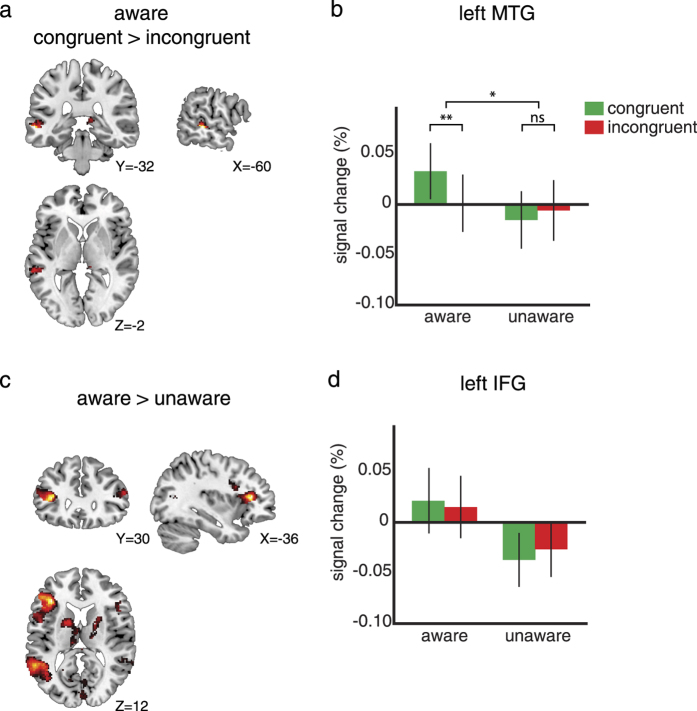
Figure 2. fMRI results.
(a) The contrast between congruent and incongruent conditions plotted on frontal, sagittal, and transversal slices of an MNI brain (p < 0.01 uncorrected for illustration purposes). The only significant modulation because of congruency is localized in lMTG (n = 23). (b) Within the lMTG ROI (based on the independent language localizer) the percentage signal change for the congruent (green) and incongruent (red) conditions is plotted for both the aware (left) and unaware (right) conditions. Only the aware condition shows a congruency effect. (c) The contrast between aware and unaware conditions shows significantly more activation in the lMTG and lIFG for the aware condition (p < 0.01 uncorrected for illustration purposes). (d) Within the lIFG region from the contrast between aware and unaware conditions, the percentage signal change for the congruent and incongruent conditions is plotted. There is no modulation of congruency for either the aware or the unaware condition.

