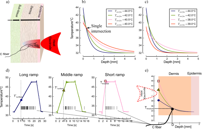
Figure 1. Experimental conditions and schematic of skin model.
(a) Schematic of skin model and a C-fiber. In a typical experimental protocol a heat electrode is placed on the surface of the skin. (b-c) Drift of different surface temperatures for all locations in the skin up to a depth of D = 5 mm as a schematic illustration. Each curve refers to a different surface temperature at different time points after stimulation onset of different experimental conditions. (b) All four curves intersect in a single point, which refers to threshold and depth of nerve endings. (c) There is no single solution of heat equation that is consistent for all four experimental conditions. (d) An example of in this study applied experimental protocol. Three heat ramped stimuli with different ramp durations were applied on the dermal side of the skin while the responses of one C-MH nociceptor to all three stimulus conditions were simultaneously recorded. The surface threshold temperatures Tthreshold are associated with the first spike times τ for all three conditions. (e) Transfer of initial Tthreshold of all conditions through the skin layers for the example neuron. The curves intersect at the location of receptor and the same threshold temperature. Note that in our experiments the heat electrode was placed on the inside of the skin.

