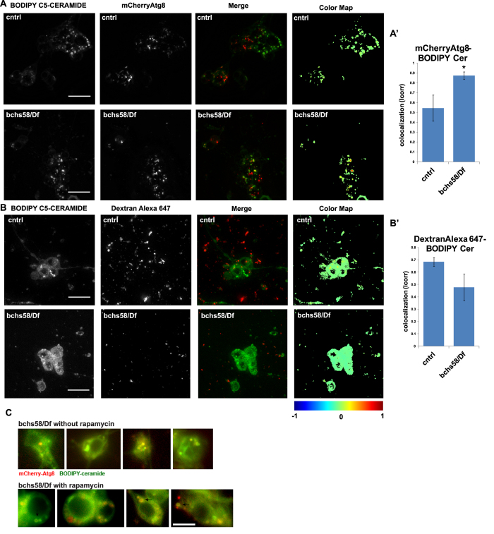
Figure 3. BODIPY-C5 Ceramide in bchs primary neurons accumulates aberrantly in mCherry-Atg8-expressing autophagosomal compartments.
Live primary neurons incubated with BODIPY C5-ceramide and imaged after chase in label-free medium, in a bchs58/Df; UAS-mCherry-Atg8 expressing background (A) and primary neurons from bchs58/Df brains treated with dextran-Alexa647 (B) to label endolysosomes. Error bars represent mean ± SEM of correlation coefficient denoting extent of co-localization between ceramide and (A) Atg8, or (B) Dextran, from four and three experiments, respectively. Scale bar = 10 μm. Co-localization analysis was carried out using the Fiji co-localization color map plug-in (https://sites.google.com/site/colocalizationcolormap/). (C) mCherry-Atg8 compartments in bchs primary neurons incubated with rapamycin (representative cells in bottom row) appear to clear BODIPY-ceramide more effectively, resulting in hollow compartments rather than the solid BODIPY-ceramide spots seen in untreated neurons (top row).

