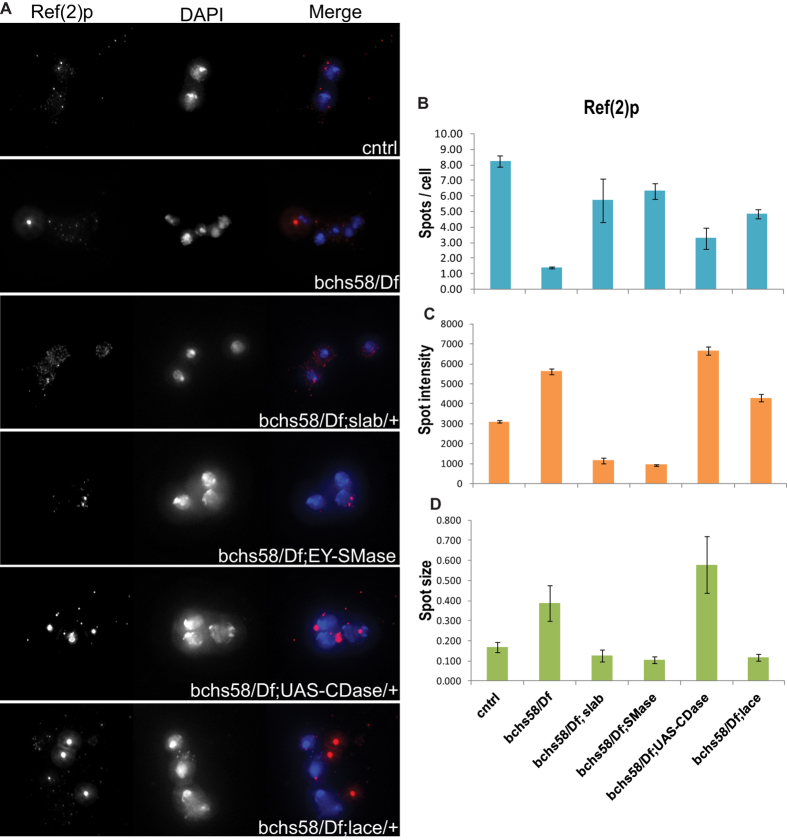
Figure 4. bchs and its modifiers affect an autophagic flux marker in primary neurons.
Primary neuron cultures of bchs58/Df and bchs58/Df larval brain in sphingolipid modifying genetic backgrounds were immunostained with Ref(2) p antibody (A, left panel) and the images were quantified for number of punctae (spots) per cell (B), spot intensity (C) and spot size (D). DAPI was used for nuclear staining. Error bars represent ± SEM.

