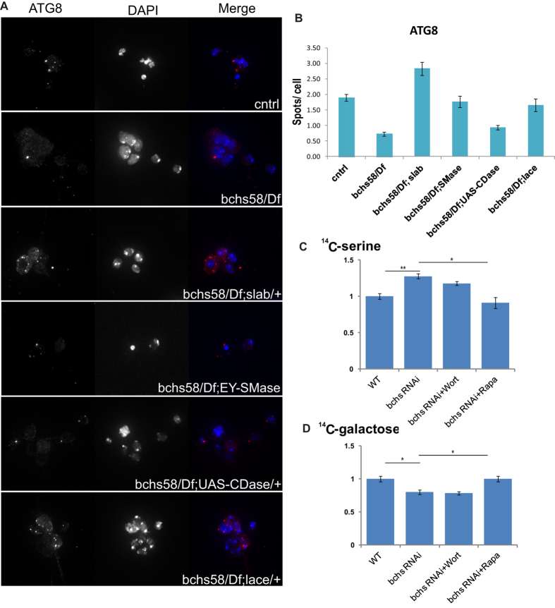
Figure 5. bchs mutants alter autophagic induction and imbalance in recycling vs. de novo sources of ceramide.
Primary neuron cultures of bchs58/Df and bchs58/Df in sphingolipid modifying genetic backgrounds were immunostained with Atg8 antibody (A, left panel) and spot number per cell was quantified similarly to Ref(2)p in Fig. 4 (B). DAPI was used for nuclear staining. (C,D) TLC quantification of total sphingolipids extracted from S2R + cells treated with bchs dsRNA and grown in medium containing either 14C-serine (C) or 14C-galactose (D) for 24 hrs. For quantification, total lipid was normalized to total protein levels and 14C signal was calculated by densitometry of the autoradiograph.

