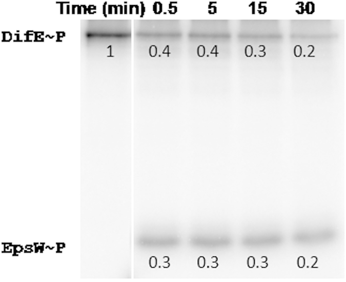
Figure 4. In vitro phosphotransfer from DifE~P to EpsW.

Prephosphorylated DifE~P labeled with [γ-32 P]ATP was mixed with equimolar amounts of EpsW and incubated for the indicated times in minutes (min). Samples were separated by SDS-PAGE and analyzed by phosphorimaging as described in Methods. The number below each band is the relative radioactivity as normalized to that of DifE~P without EpsW as loaded in the first lane. The position of each protein is indicated on the left.
