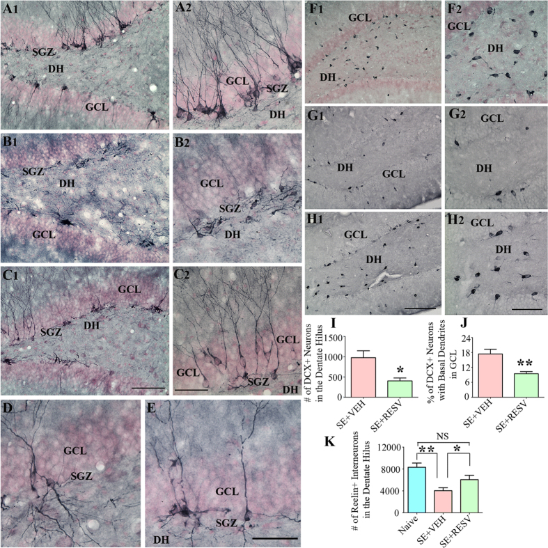
Figure 5. Resveratrol (RESV) treatment after status epilepticus (SE) curbed abnormal hippocampal neurogenesis with preservation of interneurons expressing reelin.
Figures A1, B1 and C1 show the distribution of doublecortin (DCX) positive newly born neurons in the dentate gyrus (DG) subgranular zone (SGZ), granule cell layer (GCL) and hilus (DH) from a naïve control rat (A1) and rats that received vehicle (VEH; B1) or RESV (C1) after SE. A2, B2 and C2 are magnified views of regions from A1, B1 and C1 showing the morphology of DCX + neurons. Note that, DCX + neurons are restricted to the SGZ-GCL with dendrites projecting into the molecular layer in the DG of naive control rat (A1, A2) whereas, in the DG of rat receiving VEH after SE, DCX + neurons have moved mostly into the DH (B1, B2), implying abnormal migration. In contrast, in the DG of rat receiving RESV during and after SE, majority of DCX + neurons remained in the SGZ-GCL, depicting minimal abnormal migration. Figures D and E show DCX + newly born granule cells exhibiting basal dendrites projecting into the DH in rats that received VEH (D) or RESV (E) after SE. Figures F1, G1 and H1 show the distribution of reelin + interneurons in the DH from a naïve control rat (F1) and rats that received VEH (G1) or RESV (H1) after SE. F2, G2 and H2 are magnified views of regions from F1, G1 and H1. Scale bar: A, B1, C1, F1, G1 and H1, 200 μm; A2, B2, C2, F2, G2 and H2, 100 μm; D and E, 50 μm. Bar charts in I and J compare the numbers of DCX + neurons in the DH (I) and percentages of DCX + neurons in the GCL exhibiting basal dendrites (J). Rats receiving RESV during and after SE display reduced numbers of DCX + neurons into the DH and reduced percentages of DCX + neurons exhibiting basal dendrites. Bar chart in K compares the numbers of reelin + interneurons between groups. Rats receiving VEH after SE show considerable loss of reelin + interneurons in comparison to the other two groups, whereas rats receiving RESV during and after SE demonstrate no loss of reelin + interneurons. *p < 0.05; **p < 0.01; NS, not significant.

