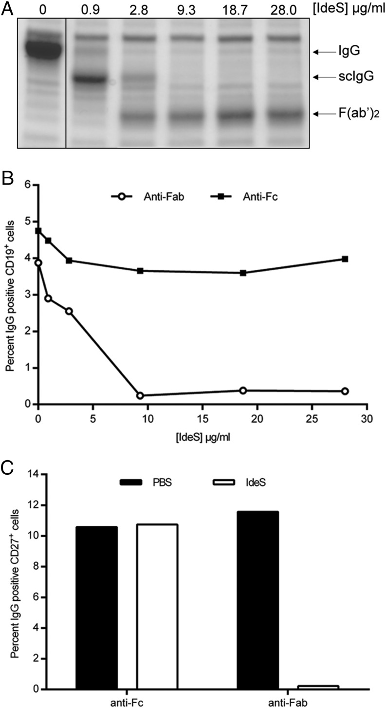
FIGURE 2.
IdeS cleaves the IgG-type of BCR with similar efficacy as soluble IgG. (A) Heparinized peripheral blood was treated with PBS or different concentrations of IdeS. After an incubation period, plasma was isolated and separated by SDS-PAGE. Intact IgG, scIgG, and F(ab′)2 fragments are indicated to the right. (B) PBMCs purified from the same PBS- or IdeS-treated blood were double stained for CD19+ (PE-coupled Ab) and anti-Fc or anti-Fab (biotin-conjugated Abs, followed by streptavidin-allophycocyanin) and analyzed by flow cytometry. Lymphocytes were gated using forward scatter/side scatter, and CD19+ cells within the lymphocyte gate were monitored for anti-Fab and anti-Fc signal. The data are representative of two independent experiments. (C) B cells were negatively selected (RosetteSep) after PBS or IdeS treatment (30 μg/ml). This CD19+-enriched population was double stained for CD27+ (PE-conjugated Ab) and anti-Fab or anti-Fc (biotin-conjugated Abs, followed by streptavidin-allophycocyanin) and analyzed by flow cytometry. The y-axis shows the percentage of CD27+ and IgG+ double-positive cells. The data are representative of two independent experiments.

