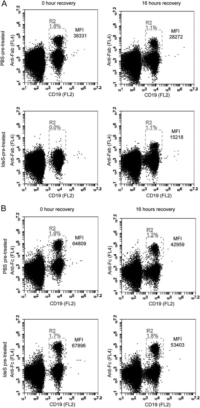
FIGURE 4.
Recovery of IgG-type BCR expression on ex vivo IdeS-treated PBMCs. PBMCs were treated with PBS or IdeS (30 μg/ml) prior to the start of staining. Lymphocytes were gated using forward scatter/side scatter (data not shown), and CD19+ cells (PE-conjugated Ab) within the lymphocyte gate were monitored for anti-Fab or anti-Fc signal using biotin-conjugated fragment-specific Abs, followed by streptavidin-allophycocyanin. (A) Flow cytometry analysis of anti-Fab signal on CD19+ cells immediately after PBS or IdeS treatment and after 16 h of IdeS-free culturing. Double-positive cells are found in R2 and expressed as the percentage of cells in the lymphocyte gate. MFI (anti-Fab) in R2 is shown. (B) Flow cytometry analysis of anti-Fc signal on CD19+ cells immediately after PBS or IdeS treatment and after 16 h of IdeS-free culturing. Double-positive cells are found in R2 and expressed as the percentage of cells in the lymphocyte gate. MFI (anti-Fc) in R2 is shown. The data are representative of two independent experiments.

