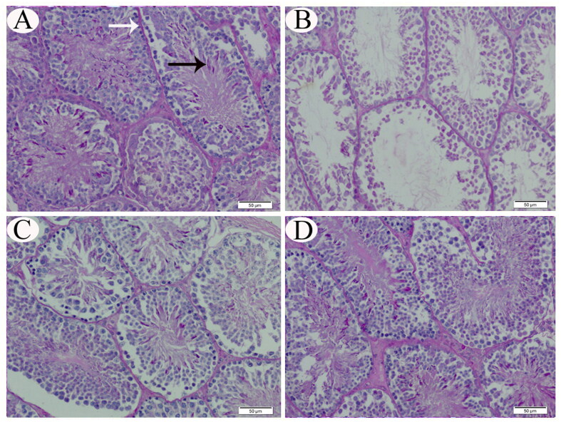
Figure 4.
Representative photomicrograph (PAS stain) of testicular tissue sections from control (A), CP-intoxicated (B), group III (CP-SES 10 mg/kg) (C), and group IV (CP-SES 20 mg/kg) rats (D). PAS-positive particles were observed in the cytoplasm of upper series cells of the germinal epithelium close to the lumen of seminiferous tubules (black arrow) and the tubular basement membrane (white arrow) of the testicular tissue sections from control and group IV rats. In contrast, PAS-positive materials were detected less in CP-intoxicated and group III rats.

