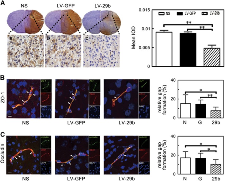
Figure 4.
IgG leakage and the breakage of zonula occludens-1 (ZO-1) and occludin were attenuated after microRNA-29b (miR-29b) overexpression. (A) Photomicrography represented IgG staining in the ischemic perifocal region in the LV-miR-29b-, LV-GFP-, and saline-treated mice after 3 days of middle cerebral artery occlusion (MCAO). Subpanels (a–c) were magnified to the box region. Scale bar=100 μm. Bar graph showed the quantification of IgG leakage. **P<0.01, data are mean±s.d., n=6 per group. (B and C) The microphotograph presented tight junction protein ZO-1 and occludin immunostaining in perifocal region after 3 days of MCAO in the LV-miR-29b-, LV-GFP-, and saline-injected groups. The arrowhead indicated the gap caused by ZO-1 and occludin protein destruction. Bar graph showed the quantification of gap length. *P<0.05; **P<0.01, data are mean±s.d., n=6 per group. G: LV-GFP; 29b: LV-miR-29b; LV, lentivirus; N: normal saline.

