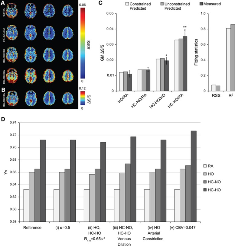
Figure 5.
Group averaged fractional signal change maps, multivariate signal change fitting, and simulated effects of changes to physiologic assumptions on Yv. (A) Fractional signal change maps for the gas pairings demonstrate hypercapnic–hyperoxia (HC-HO) interleaved with room air (HC-HO/RA) exhibits significantly elevated signal change compared with other gas stimuli. (B) Hypercapnic–hyperoxia interleaved with room air (HC-HO/RA) scaled to similar contrast as other gas pairings. (C) Measured and predicted gray matter signal change bar graphs for the three gas pairings of interest plus hyperoxia (HO) interleaved with room air (HO/RA). Error bars for the measured gray matter signal represent the standard error. For the measured values, *significantly >HC-NO/RA; **significantly >HC-HO/HO. Measured signal changes were significantly different across all stimulus pairings of interest; this excludes HO/RA for which t-tests were not calculated owing to only one stimulus block of this condition being applied (see Methods). (D) Simulated effects of changes to physiologic assumptions on Yv. Here we have systematically changed our physiologic assumptions to demonstrate the potential impact these assumptions have on Yv for the three gas stimuli: hyperoxia (HO), hypercapnic–normoxia (HC-NO), and hypercapnic–hyperoxia (HC-HO). Specifically, (i) we changed Grubb's α, which dictates the relationship between cerebral blood volume (CBV) and cerebral blood flow, from 0.38 to 0.5, (ii) we allowed for hyperoxic gases (HO and HC-HO) to also reduce R1,v, (iii) we allowed for both hypercapnic gases (HC-NO and HC-HO) to elicit venous vasodilation instead of only arterial dilation, (iv) we allowed for HO to elicit arterial vasoconstriction (0.001 mL/mL), and (v) we changed our assumption of the baseline gray matter CBV from 0.055 mL/mL to 0.047 mL/mL.

