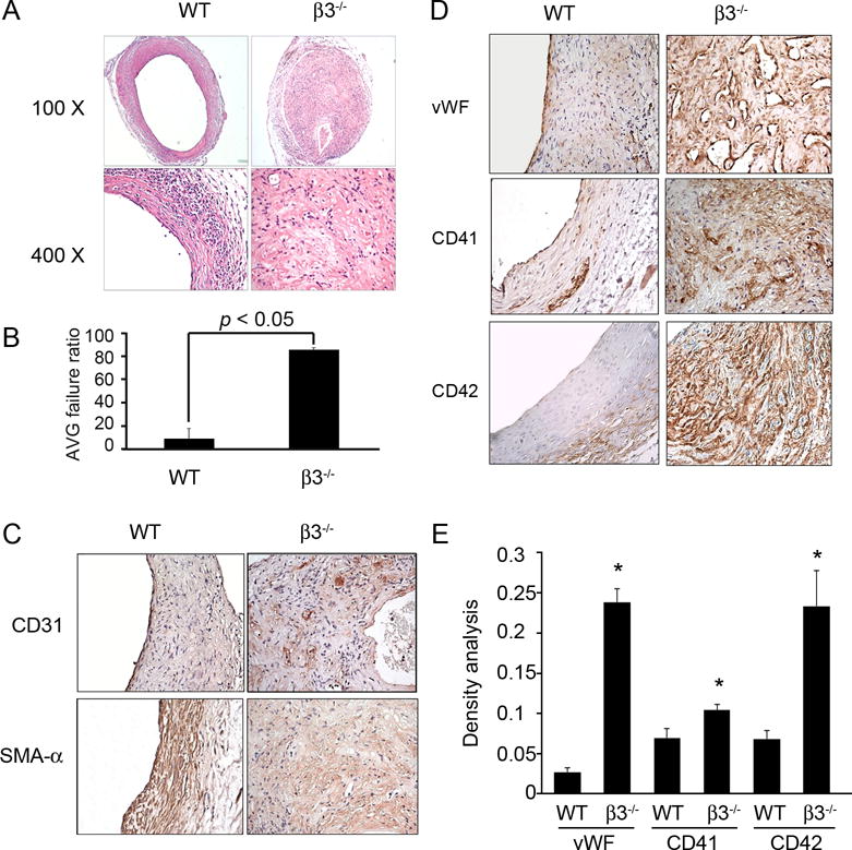
Figure 2. Integrin β3 deficiency accelerates AVG failure.

A. H & E staining of AVGs in wild type and integrin β3−/− mice show marked differences in patency. B. The ratio of failed to total AVGs was calculated. Total 15 AVGs were created in integrin β3−/− mice, and 9 in wild type mice. C and D. The difference in AVGs of wild type or integrin β3−/− mice is revealed by immunostaining with the endothelial marker, CD31 (C), smooth muscle marker SMA-α (C), and platelets markers (D). E. The density analysis of the expression of platelet markers in WT and integrin β3−/− mice (n=4).
