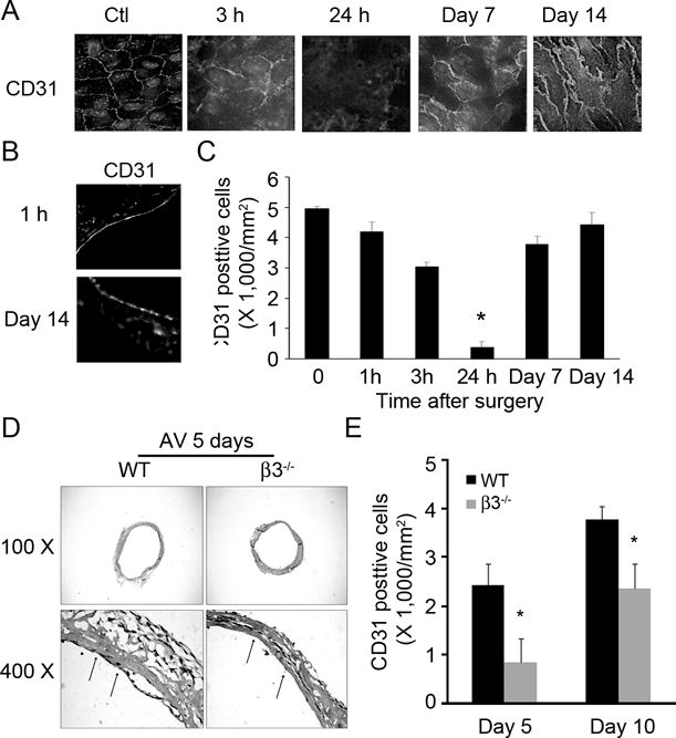
Figure 3. Integrin β3 KO delays endothelial regeneration in AVGs.

A. AVGs collected at different time points were examined by a deconvolution microscope after staining with the endothelial marker, CD31. B. Endothelial markers were present in cross sections of AVGs collected at different times from wild type mice. C. Statistical analysis of endothelial cell numbers shown in panel A. D. AVGs created in wild type and integrin β3−/− mice were collected after 5 days and examined by H & E staining. E. Statistical analysis of the endothelium in AVGs from wild type and integrin β3−/− mice after enface analysis, confirmed that the absence of integrin β3 delays regeneration. Representative data were from at least 3 mice.
