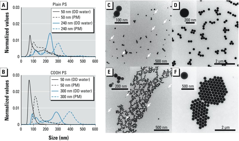
Figure 1.

Particle size distribution (A,B) and TEM analysis (C–F) of PS beads. Size distribution of plain (A) and COOH (B) PS beads was measured in DD water and PM by nanoparticle tracking analysis. (C–F) TEM micrographs of plain 50-nm (C), plain 240-nm (D), 50-nm COOH (E), and 300-nm COOH (F) PS beads in DD water. Insets show a higher magnification of the PS beads. Arrows indicate an additional fraction of smaller PS beads. Abbreviations: COOH, carboxylate-modified; DD, double distilled; PM, perfusion medium; PS, polystyrene; TEM, transmission electron microscopy.
