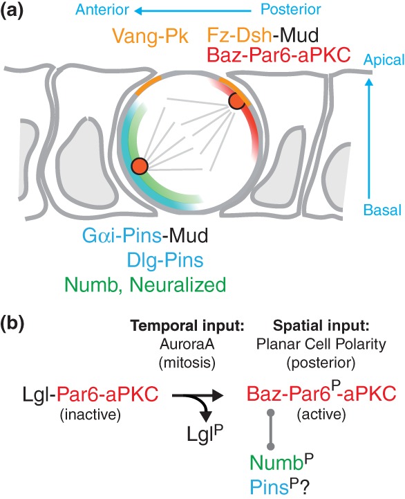
Figure 7.

Numb anterior localization and orientation of the spindle by PCP and asymmetric aPKC activity. (a) Diagram of a dividing SOP at metaphase. The posterior PCP complex includes Fz and Dsh (light orange). The Baz-Par6-aPKC complex (red) also localizes apically at the posterior cortex. Numb (green) and Pins (blue) co-localize at the anterior basal cortex. Dsh, at the posterior-apical cortex, and Pins, recruits Mud. Mud interacts with dynein and pulls on astral microtubules to line up the mitotic spindle along the anterior–posterior axis, with a slight anterior basal tilt. (b) At interphase, Lgl inhibits the active Par6-aPKC complex. Phosphorylation of Par6 by AurA leads to the aPKC-dependent release of Lgl at mitosis and to the formation of the active Baz-Par6-aPKC complex. This complex localizes at the posterior cortex in a PCP-dependent manner. This complex interacts with Numb, a target of aPKC. Phosphorylated Numb is excluded from the posterior cortex. A similar mechanism may account for the exclusion of Pins from the posterior cortex at mitosis. Thus, PCP provides a spatial input and mitosis provides a temporal input for the asymmetric localization of the Baz-Par6-aPKC complex that regulates, together with PCP, both the asymmetric localization of Numb and the orientation of the mitotic spindle.
