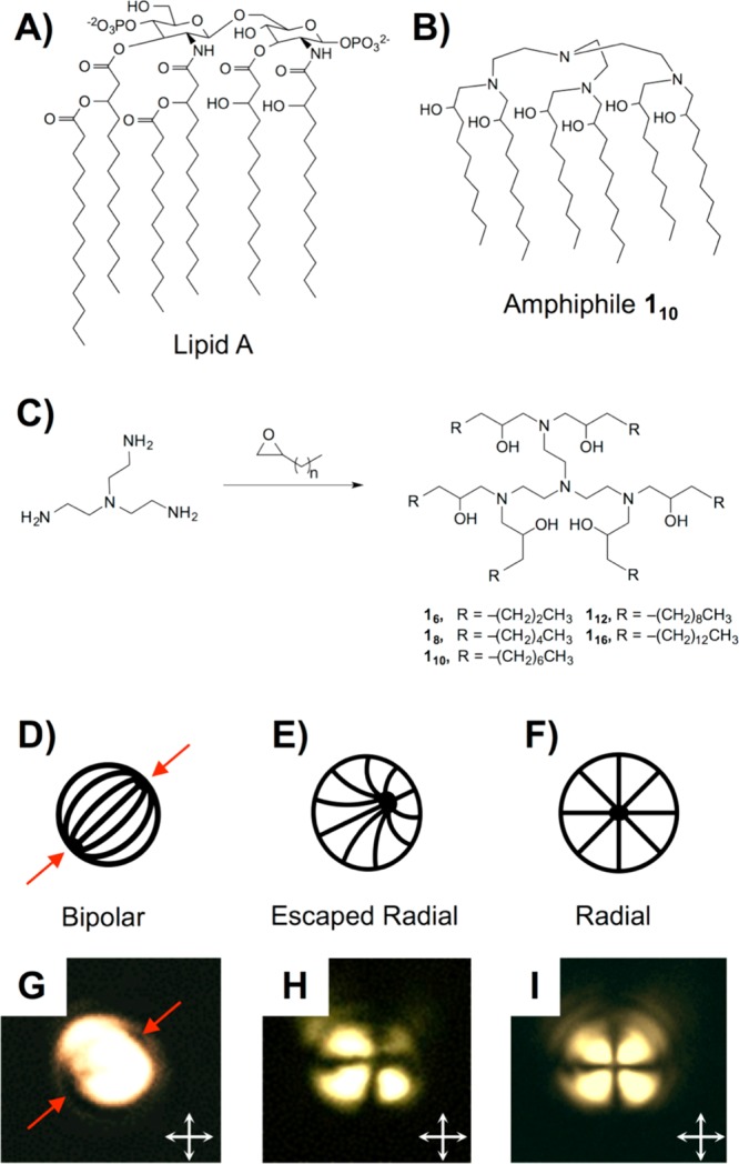
Figure 1.

(A,B) Structures of Lipid A and amphiphile 110. (C) Synthesis of six-tailed Lipid A mimics. (D–F) Director profiles for LC droplets in (D) bipolar, (E) escaped radial, and (F) radial configurations. (G–I) Polarized light micrographs of microdroplets of 5CB (diameters of ∼5 μm) prior to (G) and after (H and I) exposure to amphiphile 110 (see text). Red arrows in (D) and (G) point to topological defects in the bipolar droplets; white arrows (G–I) indicate orientation of crossed polarizers.
