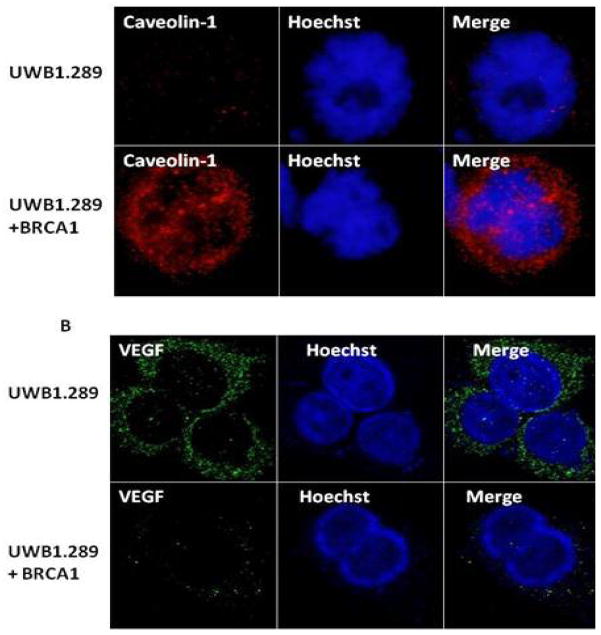
Figure 1.
Loss of Caveolin-1 and induction of VEGF expression in BRCA1 mutant HGSEOC cells UWB1.289 as detected by immunofluorescence analysis. UWB1.289 and UWB1.289 + BRCA1 cells were seeded into six-well plates. The nuclei were visualized with DNA stain Hoechst 33258. Cells were fixed in methanol and probed with A: Caveolin-1(Santa Cruz, 1/250) B: VEGF(Santa Cruz, 1/250) followed by Alexa Fluor 568/488 labeled secondary antibody(Invitrogen, 1/200) staining as described (54). The images were taken using fluorescent microscope (100X, oil Olympus)

