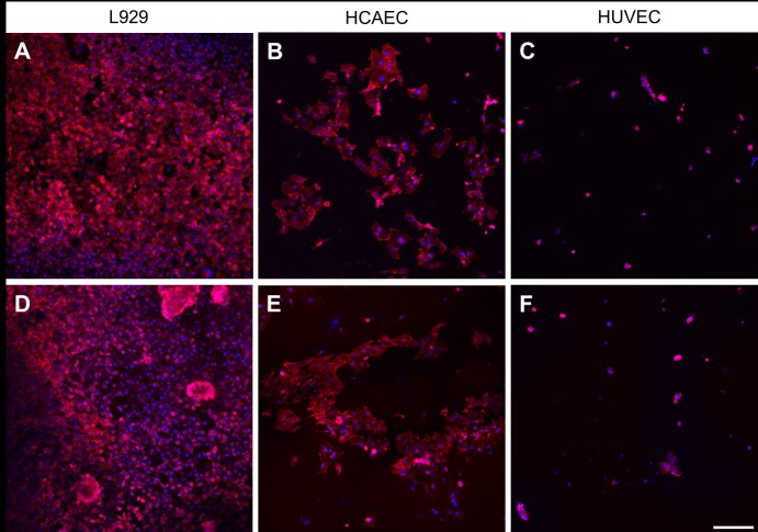
Fig 5. Confocal images of cells grown on two different polymers.
Actin filaments are seen in red (phalloidin-staining), cell nuclei in blue (Hoechst-staining) The upper row demonstrates the cell adhesion of L929 (A), HCAEC (B) and HUVEC (C) after incubation on unmodified P(LLA-co-GA).The lower row demonstrates the adhesion of the same cells, L929 (D), HCAEC (E) and HUVEC (F) following incubation on unmodified P(LLA-co-CL). Scale bar = 200 μm.

