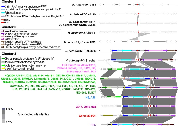
Fig 3. Genomic organization of the region encompassing hpn and hpn-2.
Conserved homologous genes are shown with similar colors, hypothetical proteins are shown in gray, while non-conserved genes are in white. Hpn (in red) is located in-between two conserved cluster of genes (clusters 1 and 2), while hpn-2 is located either near hpn on the same strand or on the opposite side of cluster 1 on the opposite strand. Helicobacter strains harboring similar genomic organization are listed on the left with similar colors.

